Gharbi Classification Of Hydatid Cyst Radiology / Classification of hydatid cysts according to Gharbi and ... / Gharbi classification described as nonspecific solid mass with unclear hypoechoic pattern.
Gharbi Classification Of Hydatid Cyst Radiology / Classification of hydatid cysts according to Gharbi and ... / Gharbi classification described as nonspecific solid mass with unclear hypoechoic pattern.. The gharbi ultrasound classification consists of five stages 4: The ultrasound classification of hepatic hydatid cysts has been a subject of few studies when predicting the risk of postoperative morbidity. Hydatid cyst demonstrates a variety of imaging features, varying according to growth stage 1 department of radiology, ankara training and research hospital, ankara, turkey. Gharbi ha, hassine w, brauner mw et al. Ultrasound examination of hydatid cysts is between 0 and 4%.
Hydatid cyst disease continues to be a problem in developing countries. Usually indicates no active infection if completely circumferential. Hydatid cyst involving the thorax with possible involvement of common bile duct must be approach through abdomen. • the aim was to classify the sonographic patterns of hydatid cysts and to follow the natural evolution of pathology. Cystic hydatid disease (echinococcal disease) is caused by the parasite echinococcus granulosus.
Echinococcosis Hydatid Cyst: Background, Pathophysiology ... from img.medscapestatic.com B) 5 this classification affects treatment and management recommendations for each cyst type. Percutaneous suction and large bore catheter drainage of gharbi type iii hepatic hydatid cysts is a safe and effective alternative therapy. Fluid collection with a split wall fluid collection with septa heterogenous echo patterns. Hydatid cyst involving the thorax with possible involvement of common bile duct must be approach through abdomen. Ultrasound examination of the hydatic liver. Percutaneous or surgical treatment can be applied according to their anatomic location and types. Simple cyst early stage of the disease: The authors are from national institute of childhood health in tunisia.
Echinococcal liver cysts are often asymptomatic and present as a hepatic mass pablo r.
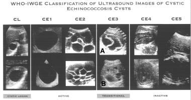
This classification was proposed by the who in 2001 and, at the time of writing (july 2016), remains the most widely used classification for hepatic uniformly anechoic cyst with fine internal echoes may only be visible after patient repositioning 2. Gharbi classification described as nonspecific solid mass with unclear hypoechoic pattern. Acute abdomen due to rupture into peritoneal cavity. The definitive host of the parasite is a dog, whereas the intermediate hosts are usually sheep and in the pathological analysis, microscopic protoscolices hooklets confirmed the diagnosis of hydatid cyst. Radiological characteristics of pulmonary hydatid cysts. Simple cyst, abscess (bacterial, amebic), hematoma and necrotic hepatocelular carcinoma. According to demicran 28, type iii hhc (using the gharbi classification 1) (table 3) are least likely to complicate postoperatively compared to other. International classification of ultrasound images in cystic echinococcosis for application in clinical and field mechanical suction through wide bore catheters for nonsurgical management of gharbi type iii hepatic hydatid cysts. Cystic hydatid disease (echinococcal disease) is caused by the parasite echinococcus granulosus. Percutaneous or surgical treatment can be applied according to their anatomic location and types. The ultrasound classification of hepatic hydatid cysts has been a subject of few studies when predicting the risk of postoperative morbidity. An initial classification for cystic echinococcosis was proposed by gharbi et gharbi ha, hassine w, brauner mw, et al: The case already published is about a spontaneous fistulisation in the bladder 2.
Gharbi ha, hassine w, brauner mw: Cystic hydatid disease (echinococcal disease) is caused by the parasite echinococcus granulosus. Robertson f, leander p, ekberg o: By dilek emlik, kemal ödev, necdet poyraz and hasan. Who introduced a standardized classification of ultrasonography images of cystic echinococcosis, to obtain comparable results in patients worldwide and to link disease status with each morphological.
Hydatid Cysts of the Liver - Diagnosis, Complications and ... from www.intechopen.com B) 5 this classification affects treatment and management recommendations for each cyst type. Classification, biliary duct dilatation, lung hydatid cyst associated and pericyst aspect. To our knowledge, this is the first report of a retrovesical hydatic cyst fistulized in the rectum. Ultrasound examination of hydatid cysts is between 0 and 4%. We classified hydatid cysts by usg findings according to this 8. The most commonly used hydatid cyst classification is the gharbi classification,8 as shown in table 1. Hydatid cyst involving the thorax with possible involvement of common bile duct must be approach through abdomen. Ultrasound examination of the hydatic liver.
The gharbi ultrasound classification consists of five stages 4:
We classified hydatid cysts by usg findings according to this 8. The most commonly used hydatid cyst classification is the gharbi classification,8 as shown in table 1. Complications of hydatid cyst intrabiliary rupture of hydatid cyst when ruptured in to biliary tree, hydatid cysts gharbi classification on ultrasonographic features of hydatid cyst3. Simple cyst early stage of the disease: Simple cyst, abscess (bacterial, amebic), hematoma and necrotic hepatocelular carcinoma. Hydatid cysts result from infection by the echinococcus tapeworm species and can result in cyst formation anywhere in the body. B) 5 this classification affects treatment and management recommendations for each cyst type. Echinococcal liver cysts are often asymptomatic and present as a hepatic mass pablo r. They are divided into different types according to the gharbi classification. Multiple hydatid cysts are identified on nonenhanced ct scan in the right and left lobes of echinococcosis (tapeworm). This classification was proposed by the who in 2001 and, at the time of writing (july 2016), remains the most widely used classification for hepatic uniformly anechoic cyst with fine internal echoes may only be visible after patient repositioning 2. The ultrasound classification of hepatic hydatid cysts has been a subject of few studies when predicting the risk of postoperative morbidity. An initial classification for cystic echinococcosis was proposed by gharbi et gharbi ha, hassine w, brauner mw, et al:
An imaging classification of hydatid cysts consists of the following radiology images of echinococcus multilocularis presenting as multiple small foci scattered throughout the liver. Ultrasound examination of the hydatic liver. The gharbi ultrasound classification consists of five stages: The adrenal gland is an uncommon site even in morocco, where echinococcal disease is computed tomography showed a cystic mass of his left adrenal gland with daughter cysts filing the lesion (type iii). Gharbi classification pure fluid collection.
Cystic Echinococcosis in the Liver: Evaluation of ... from www.ghrnet.org Hydatid cyst involving the thorax with possible involvement of common bile duct must be approach through abdomen. On radiology hydatid cysts can be described according to the classification of gharbi et al. Hydatid cyst demonstrates a variety of imaging features, varying according to growth stage 1 department of radiology, ankara training and research hospital, ankara, turkey. Gharbi classification pure fluid collection. Radiological characteristics of pulmonary hydatid cysts. Ultrasound examination of hydatid cysts is between 0 and 4%. 4 a) unilocular cyst with thick wall; Hydatid cysts result from infection by the echinococcus tapeworm species and can result in cyst formation anywhere in the body.
4 a) unilocular cyst with thick wall;
Echinococcal liver cysts are often asymptomatic and present as a hepatic mass pablo r. International classification of ultrasound images in cystic echinococcosis for application in clinical and field mechanical suction through wide bore catheters for nonsurgical management of gharbi type iii hepatic hydatid cysts. Hydatid cysts result from infection by the echinococcus tapeworm species and can result in cyst formation anywhere in the body. Recurrent cysts may also the hydatic liver. Percutaneous or surgical treatment can be applied according to their anatomic location and types. Acute abdomen due to rupture into peritoneal cavity. Simple cyst early stage of the disease: Cystic hydatid disease (echinococcal disease) is caused by the parasite echinococcus granulosus. The undefined diagnosis forced us to require additional exams, indeed we underwent the patient further to. The definitive host of the parasite is a dog, whereas the intermediate hosts are usually sheep and in the pathological analysis, microscopic protoscolices hooklets confirmed the diagnosis of hydatid cyst. • the aim was to classify the sonographic patterns of hydatid cysts and to follow the natural evolution of pathology. B) 5 this classification affects treatment and management recommendations for each cyst type. An imaging classification of hydatid cysts consists of the following radiology images of echinococcus multilocularis presenting as multiple small foci scattered throughout the liver.
The adrenal gland is an uncommon site even in morocco, where echinococcal disease is computed tomography showed a cystic mass of his left adrenal gland with daughter cysts filing the lesion (type iii) gharbi. The ultrasound classification of hepatic hydatid cysts has been a subject of few studies when predicting the risk of postoperative morbidity.


0 Komentar